Systemic Right Ventricle – Transposition of the Great Arteries
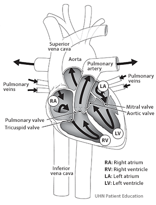
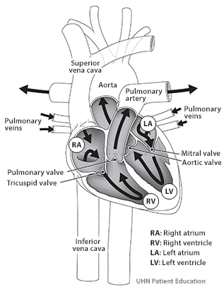
In the usual arrangement of the heart, de-oxygenated blood returning from the body through the heart comes to the right side of the heart, specifically the right atrium. From the right atrium, the blood gets directed to the lungs where it gets oxygenated. Once oxygenated, the blood gets to the left side of the heart (left atrium first and then left ventricle) and then it is pumped back to the body. This type of circulation is called biventricular circulation (image 1)
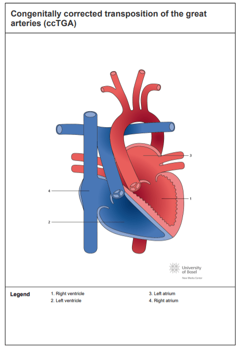
In some congenital heart diseases this right-left arrangement is inversed from birth or has had to be inversed through surgical correction. In these cases, the left ventricle will pump the blood to the lungs and the right ventricle to the body and this type of circulation is the one we refer to as a systemic right ventricle.
Systemic right ventricle are found:
- At birth in people with congenitally corrected transposition of the great arteries (ccTGA). (image 2). https://www.chd-diagrams.com/heart-disease/137
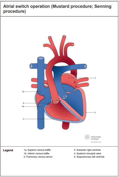
- After surgery as a result of the atrial switch surgery (Mustard or Senning) historically performed to people with transposition of the great arteries (TGA). (Image 3). https://www.chd-diagr ams.com/heart-operation/172
In the systemic right ventricle circulation the right ventricle functions as the left ventricle, pumping blood to the body. The main challenge the right ventricle faces is that the pressure in the body is up to 4 times higher than in the lungs, and the right ventricle is not naturally prepared to work against high pressures and so it must adapt when it does. The systemic right ventricle working against higher pressures gets thicker (as the pectorals with push-ups) and gets larger in volume. These two ways of adapting, although not perfect, are quite efficient and can last for decades unless affected by other cardiac issues (e.g., valve leaking, obstruction, arrhythmias).
Recent studies suggest that regular exercise, specially aerobic, contributes to improve the function of the heart and also can help reduce the pressure the systemic right ventricle has to face by reducing blood pressure, obesity and improving other cardiovascular diseases.
Normal Circulation
In the usual arrangement of the heart, de-oxygenated blood returning from the body through the heart comes to the right side of the heart, specifically the right atrium. From the right atrium, the blood gets directed to the lungs where it gets oxygenated. Once oxygenated, the blood gets to the left side of the heart (left atrium first and then left ventricle) and then it is pumped back to the body. This type of circulation is called biventricular circulation.
Fontan Circulation
Among the broad spectrum of congenital heart diseases there are many that have only a single ventricle supporting both circulations: meaning pumping the blood to the lungs and the body. In this case, de-oxygenated blood and oxygenated blood both arrives in that single ventricle. As a result the level of oxygen in the blood that is pumped into the body is lower than what expected and the single ventricle receives to a higher volume of blood than in a biventricular circulation, increasing its work. Unfortunately the single ventricle cannot sustain this type of work for long time.
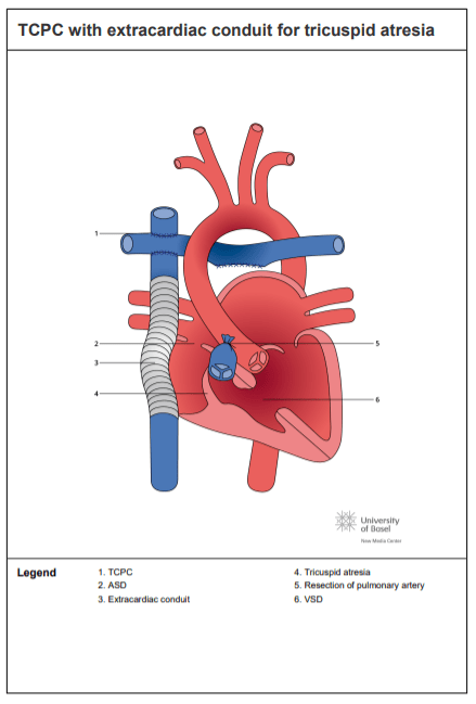
In the late 1960’s, Dr. Fontan performed the first surgical procedure to avoid deoxygenated blood arriving to the body and oxygenated blood arriving from the lungs to mix in the single ventricle. This intervention is now known as ‘Fontan procedure’ and although there have been multiple modifications of the original operation, the main idea of the ‘Fontan circulation’ remain the same: mainly, the deoxygenated blood returning from the body is directed straight to the lungs without the help of a ventricle (see image) and the single ventricle pumps the blood to the body. The surgery is usually performed in two or three steps during childhood, meaning that you will have probably gone under cardiac surgery multiple times until the Fontan circulation was completed.
In the Fontan circulation, the quantity of blood arriving to the lungs and afterwards to the heart is less than in a biventricular circulation because there is no ventricle pushing the blood into the lungs. The main force driving the blood to the lung is the difference in pressure between the venous system and the pulmonary circulation. In other words, the lungs to the blood are the same as a highway car accident to the traffic: there is congestion upstream and restricted flow downstream. At rest this situation is usually well balanced, whereas during exercise,the downstream flow of blood from the lungs to the venous system tends to increase and some patients might experience some degree of exercise intolerance, particularly with activities of high intensity.
Nevertheless recent studies suggest that regular exercise contributes to improve the function of the pulmonary circulation and the heart in the setting of a Fontan circuit, as you will read in the next learning module.
